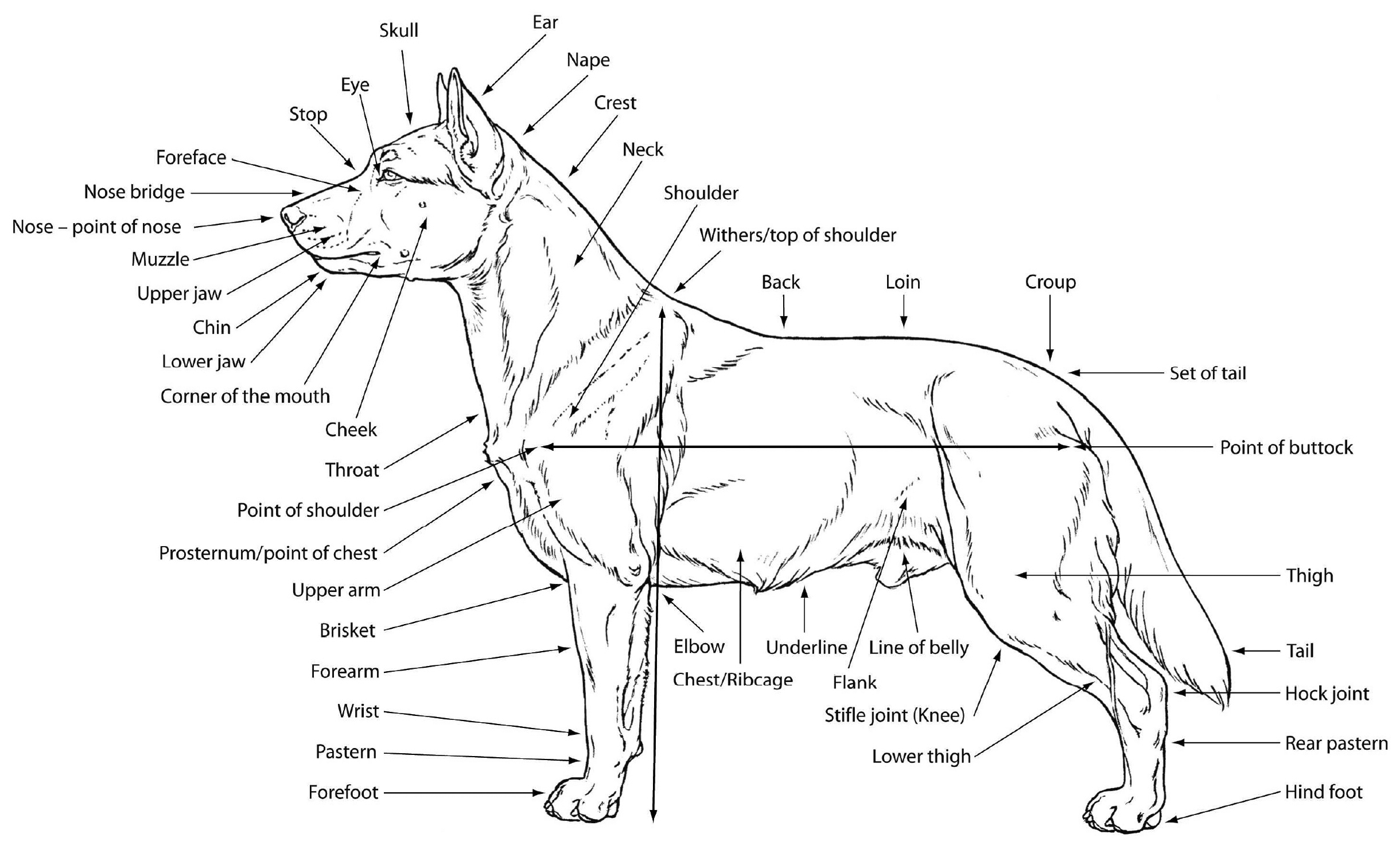
Labeled images in the transverse plane of a healthy dog’s whole body, using tomodensitometry. Learn how many teeth dogs have and what to do if they're missing or broken. Yet he is an evolutionary relative of the horse and so will forever be compared with the horse. Here are presented scientific illustrations of the canine muscles and skeleton from different anatomical standard views (lateral, medial, cranial, caudal, dorsal, ventral / palmar.). Dogs have four types of teeth, each varying in function, location in.
This ligament is like the anterior cruciate ligament (acl) in humans. If this plane were in the midline of the body, this is the median plane or median sagittal plane. Muscle, organ and skeletal anatomy). Web understand dog anatomy with our canine charts and models, including skeletons and pathology models. Here are presented scientific illustrations of the canine muscles and skeleton from different anatomical standard views (lateral, medial, cranial, caudal, dorsal, ventral / palmar.).
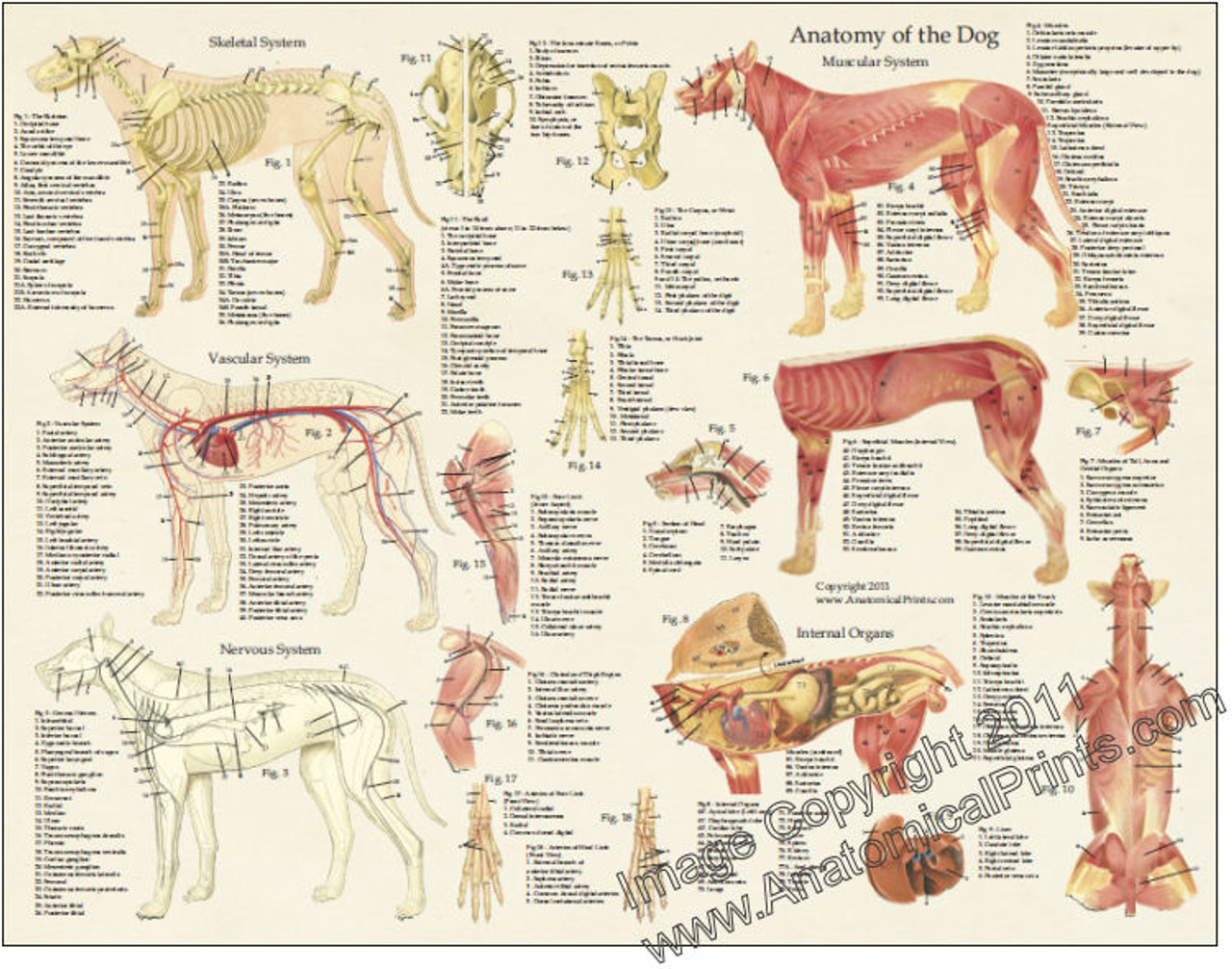
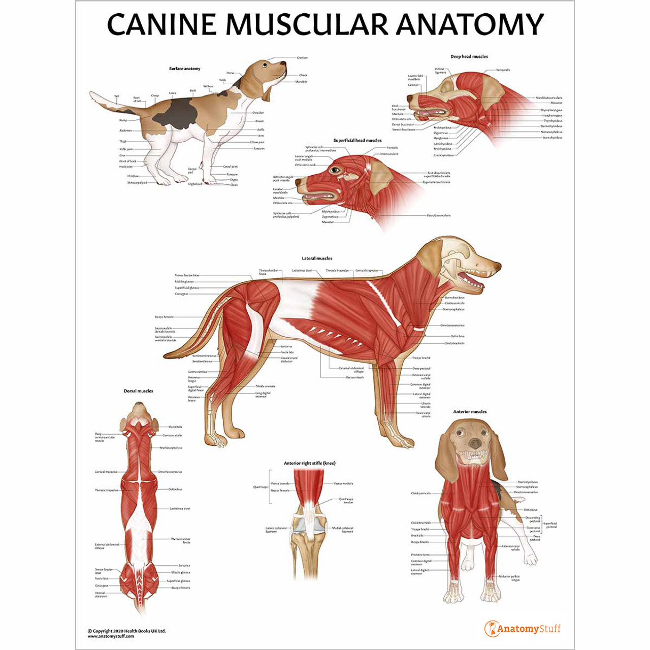
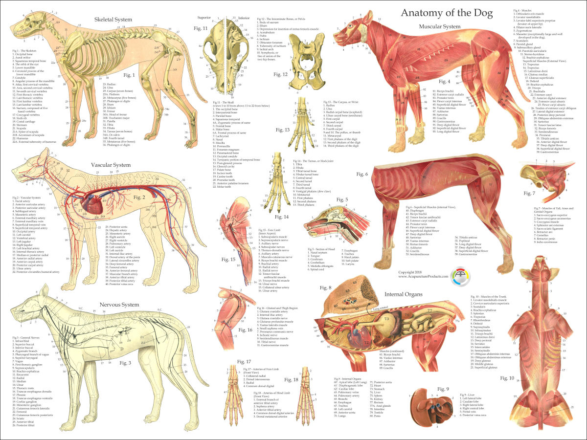
Web the anatomy of a dog includes its skeletal structure, reproductive system, the internal organs, and its external appearance. Web this veterinary anatomy module contains 608 illustrations on the canine myology. The first two web based reviews involve identifying normal airways of the canine thorax on a right lateral and on a ventrodorsal radiograph. Robin downing, dvm, daapm, dacvsmr, cvpp, crpp. Web three of our most popular anatomically accurate charts of the skeleton, musculature and internal organs of a dog.
Canine Internal Anatomy Poster Dog Organs Laminated Chart

Canine Skeleton Poster Clinical Charts and Supplies
Canine Dental Anatomy Chart Dog Teeth Jaw Poster
Dog Anatomical Chart Bones and Muscles
Dog Anatomy Laminated Poster Clinical Charts and Supplies
M. Douglas Wray Dog Anatomy
Canine Muscular Anatomy Chart Dog Muscles Poster Laminated
Dog Anatomy Poster
Canine Anatomy, Complete Set of 3 Charts. Buy The Set and Save! Amazon

Canine Skeletal Anatomy Laminated Chart Dog Skeleton Poster
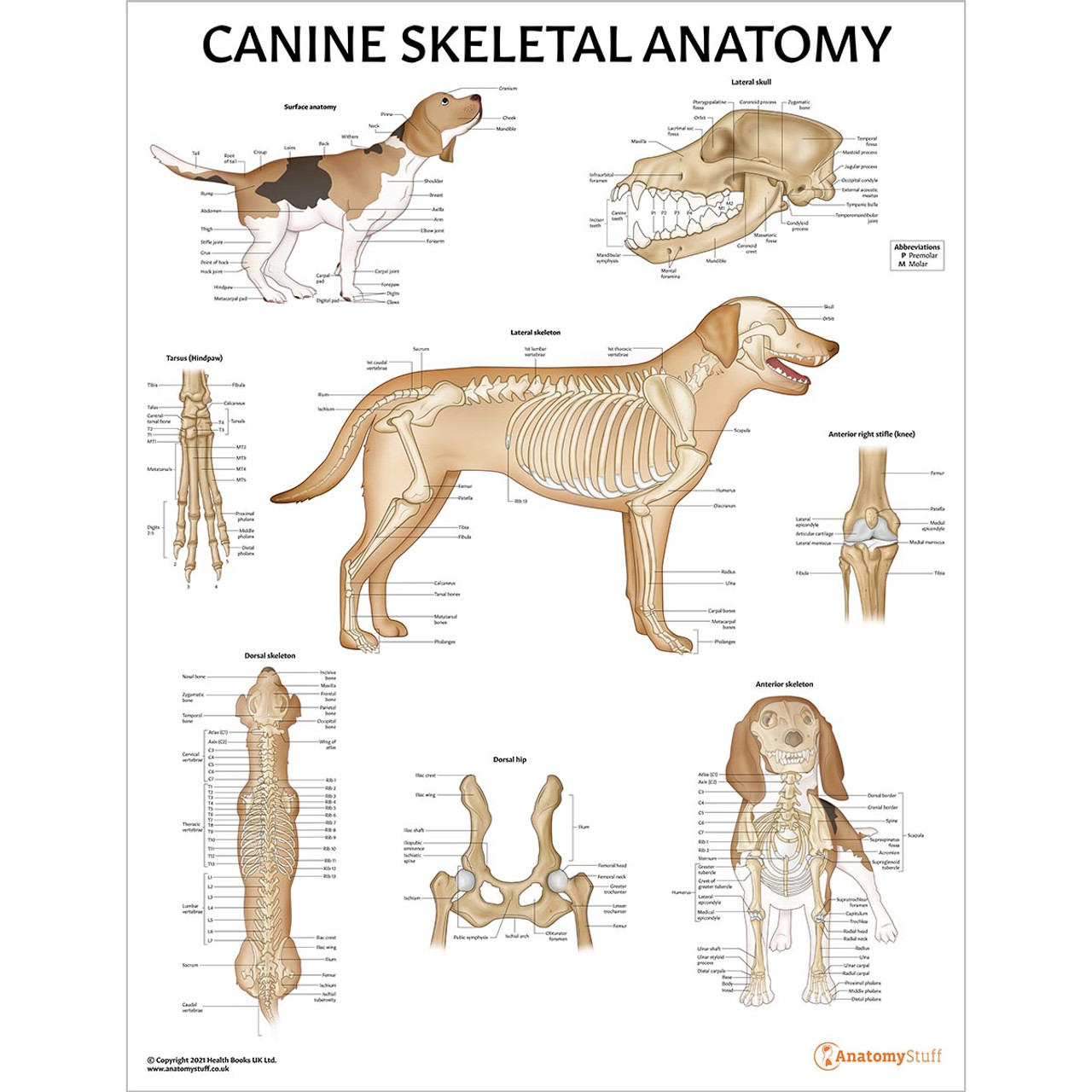
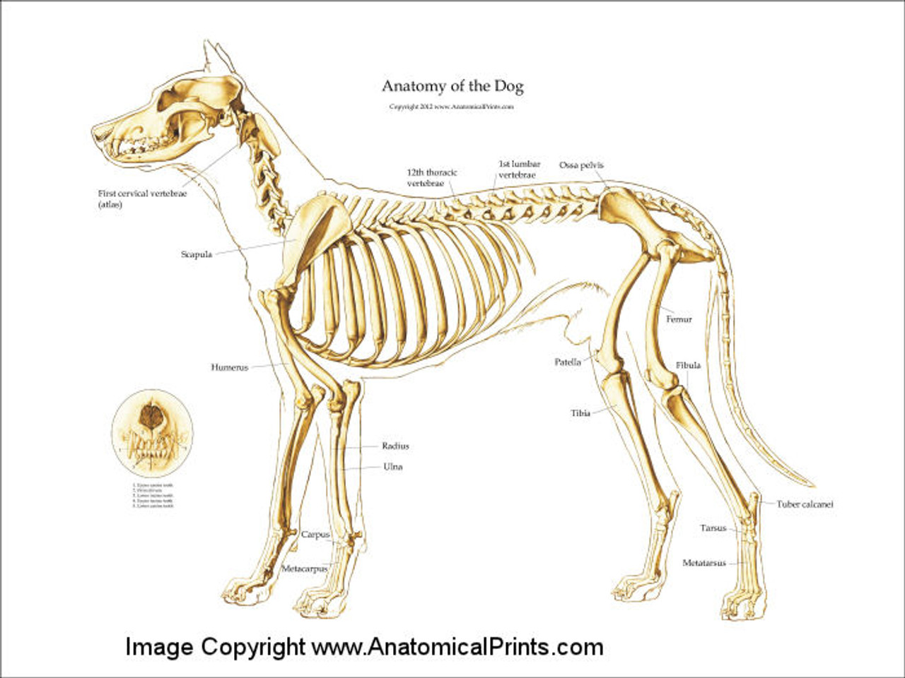
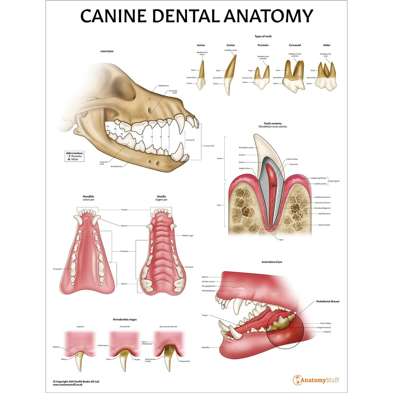
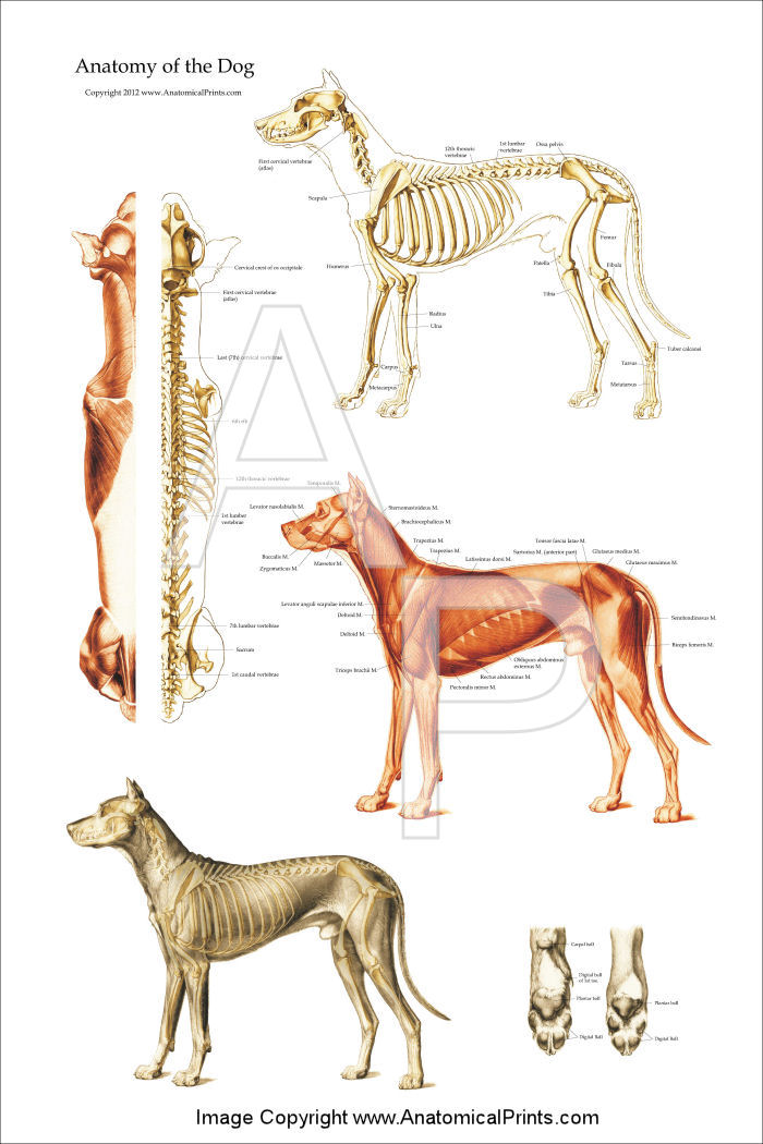
Here are presented scientific illustrations of the canine muscles and skeleton from different anatomical standard views (lateral, medial, cranial, caudal, dorsal, ventral / palmar.). They have a heart and circulatory system to transport blood, lungs to take in oxygen and rid the body of carbon dioxide, a digestive tract to. Web anatomy atlas of the canine general anatomy: A basic understanding of dental anatomy will help you understand dog dental charts better. One of the most common injuries to the knee in dogs is tearing of the cranial cruciate ligament (ccl). Web here are presented scientific illustrations of the canine skeleton, containing the main joints of the dog and its structures from different standard anatomical views (cranial, caudal, lateral, medial, dorsal, palmar.). Web three of our most popular anatomically accurate charts of the skeleton, musculature and internal organs of a dog. • the dorsal plane divides the dog into ventral and dorsal portions. Web by malcolm weir, dvm, msc, mph; The apex of the mandibular canine tooth lies lingual to the mental foramen and occupies a large portion of the mandible. The first two web based reviews involve identifying normal airways of the canine thorax on a right lateral and on a ventrodorsal radiograph. Muscle, organ and skeletal anatomy). Yet he is an evolutionary relative of the horse and so will forever be compared with the horse. Each diagram is meticulously labeled with little additional text since the book truly takes a pictorial approach to the topic. • the sagittal plane divides the dog into right and left portions.
This Section Will Continue To Be Updated With More Anatomy Sections In Time.
They have a heart and circulatory system to transport blood, lungs to take in oxygen and rid the body of carbon dioxide, a digestive tract to. Here are presented scientific illustrations of the canine muscles and skeleton from different anatomical standard views (lateral, medial, cranial, caudal, dorsal, ventral / palmar.). If this plane were in the midline of the body, this is the median plane or median sagittal plane. A basic understanding of dental anatomy will help you understand dog dental charts better.
There Is Only A Thin Plate Of Bone Between The Root Of The Maxillary Canine Tooth And The Nasal Cavity, Therefore This Is A Common Location For Oronasal Fistulation.
A pictorial approach by peter c. Web although dogs look very different from people, they share many of our body’s characteristics. Web here are presented scientific illustrations of the canine skeleton, containing the main joints of the dog and its structures from different standard anatomical views (cranial, caudal, lateral, medial, dorsal, palmar.). Web this veterinary anatomy module contains 608 illustrations on the canine myology.
Web Buy Veterinary Anatomy Posters And Anatomical Charts.
Web understand dog anatomy with our canine charts and models, including skeletons and pathology models. This ligament is like the anterior cruciate ligament (acl) in humans. Fully labeled illustrations and diagrams of the dog (skeleton, bones, muscles, joints, viscera, respiratory system, cardiovascular system). Web three of our most popular anatomically accurate charts of the skeleton, musculature and internal organs of a dog.
Web Dog Anatomy Details The Various Structures Of Canines (E.g.
Labeled images in the transverse plane of a healthy dog’s whole body, using tomodensitometry. The first two web based reviews involve identifying normal airways of the canine thorax on a right lateral and on a ventrodorsal radiograph. The mule, however, is still a scientific enigma. This is why animalwised brings you this dog anatomy guide where we look at the general categories for muscles, bones and organs of dogs.